This may be bad to admit, but I love a good TikTok scroll. It’s probably not the best use of my time, but I’ve come across some interesting things! New music artists to listen to, events around my city, and even some science accounts. A bit ago, I found a medical student who makes histological artwork and I was absolutely fascinated. Her name is Kait and her paintings are absolutely beautiful (@createbykait on TikTok if you are curious)!
Of course then I had to do a deep dive about histology. What actually is it? What is she really painting? I assumed it was cells from the look of it, but that was the extent of my knowledge. Turns out there is a lot more to histology than just a pretty picture.
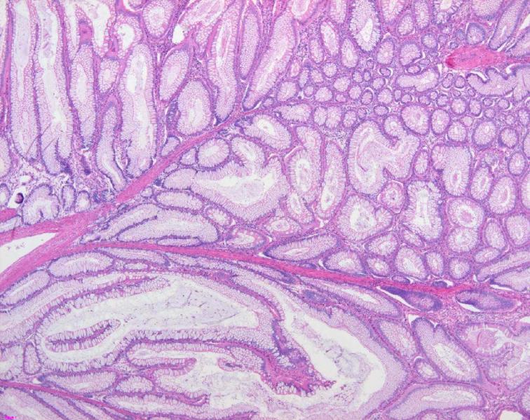
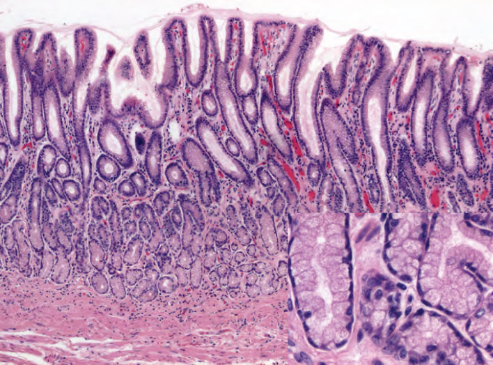
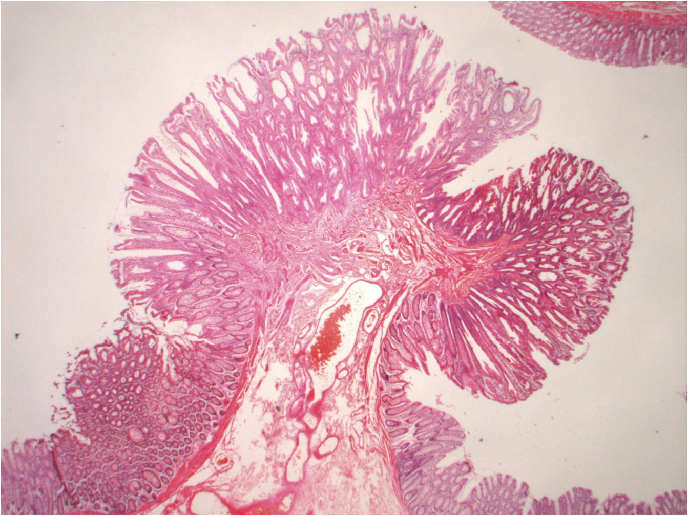
Histology is the study of microscopic structures of tissues. It usually includes sectioning a thin slice of tissue, staining it with dyes that adhere to different types of structures, then observing the slice under a microscope (NCBI Bookshelf, 2023). Histology is also the foundation for pathology, which is the study of the effects of disease on the body. I had heard pathology as a medical specialty, but again, did not know what they did.
Turns out, pathologists look at tissue samples from biopsies or autopsies to make diagnoses. I got to watch a brain biopsy earlier this year while shadowing a neurosurgical oncologist. These patients had gotten MRIs that showed a spot of something irregular, called a lesion. During the biopsy, the surgeons took a small sample of the lesion tissue to send to a pathologist to determine what it was, whether it was cancer, an aneurysm, diseased tissue, or even a parasite. The first sample they took is called the frozen section, which they would send to pathology immediately. The pathologists would quickly freeze the sample and examine it to make sure it was usable. The surgeons would have to stay scrubbed in and wait in the operating room until they got confirmation from the pathologist that the sample was good. If it wasn’t, the surgeons would go in and get another sample before closing the wound.
Pathologists also examine autopsies, or tissue from dead organisms. This is often used when there is an investigation with an unclear cause of death. This led me to Marianne Hamel, a forensic pathologist, who recognized how paradoxically beautiful these histological images can be. So much so that she created a collection of histological images that is now on display at Rudger’s University (Hamel & Johnson, n.d.). She said in an interview that she hopes to educate people about her career field, but also allow people to appreciate the beauty of the human body (Chandler, 2023).
Histology has evolved a lot since its beginnings. Hamel said photos used to be shot in black and white, making interpreting the images much more difficult. Now there is a plethora of different dyes used to make multicolored images. This offers so much more information for pathologists to gather, but also a higher level of difficulty. Learning to read [histological] slides, Hammel says, is like learning another language (Chandler, 2023).
It’s funny how what seems like just a pretty picture is actually such an intricate science and can show how beautiful life can be.
References
National Center for Biotechnology Information. (2023). NCBI Bookshelf. U.S. National Library of Medicine. https://www.ncbi.nlm.nih.gov/books/NBK557663/
Hamel, M., & Johnson, N. (n.d.). Death under glass. https://www.deathunderglass.com/
Chandler, R. (2023, October 31). Post-mortem histology artwork brings death to life. Evident Scientific. https://evidentscientific.com/en/insights/post-mortem-histology-artwork