This semester, I started doing animal work in a research lab that uses mice as model organisms. Every time I’ve explained my training to other people, I usually get a shocked response. And I know this isn’t a common thing people do, so I wanted to give y’all a look into what it’s like working in an animal lab.
The lab I work in does research using a technique called optogenetics. Opto- means light, and genetics refers to the DNA that is changed. This might sound straight out of a sci-fi novel, but by infecting a certain brain area of healthy mice with a special virus (this doesn’t make the mice sick, by the way), the virus can insert a list of instructions written in DNA into the mouse’s own DNA. These instructions tell the cells to start producing light-sensitive proteins in their membranes.
Now, this has a very important application in neuroscience. Neurons, the unit cell of the brain, fire tiny electrical signals called action potentials. If a neuron has light sensitive proteins that respond to blue light (channelrhodopsin), it fires more often. If it has light sensitive proteins that respond to yellow light (halorhodopsin), it fires less often. Using optogenetics, you can directly measure how activation of a brain area leads to behavior in real time.
So how does that factor into what I do? So far, I’ve worked on an often-overlooked but important step: collecting samples. After you inject the virus into the brain, there’s no way to check in the live animal that you did it right. You might have put the virus in the wrong place, or the virus never got its instructions to the cells, or the equipment might be broken. The only way to know for sure that your procedure worked is to take samples of the brain, and look at them under the microscope.
To do that, we first need to collect the brains. Through a process called perfusion, the brain gets fixed in paraformaldehyde (PFA), a safer version of the formaldehyde you might remember from dissections in biology class. During drop fixing, where the brain is severed from the spinal cord and removed from the head, we store it in a test tube with more PFA.
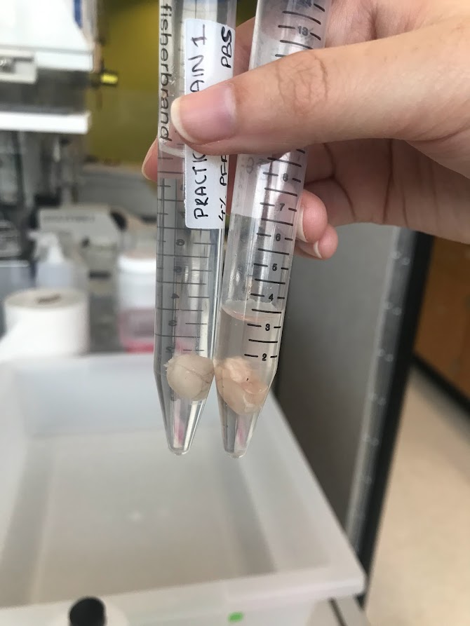
After 48 hours, the brain is ready to be sliced. There’s a lot of ways to take brain slices, but we use a vibratome. First, we locate the area of the brain that we’re trying to reach. This is where a mouse atlas comes in handy. Using a small, vibrating blade, we make slices of 50 microns–or around the width of a human hair! These slices are then stained with dyes like DAPI (which makes neurons look blue) and placed on microscope slides.
Finally, after all this work, we can see the fruits of our labor. In our lab, we use a confocal microscope because this type of microscope can show us thin layers of cells within thick layers of tissue. Using different colors of light, we can also check how well the light sensitive proteins were able to grow in the area of the brain we’re interested in. In the picture below, you can see the first picture that I took myself from a brain I perfused and sliced. There’s no injection in this brain, so it’s as close to a “natural” brain slice that you can get. The blue color is from the DAPI dye, which indicates the location of DNA in a nucleus. So, each tiny blue dot is a neuron.
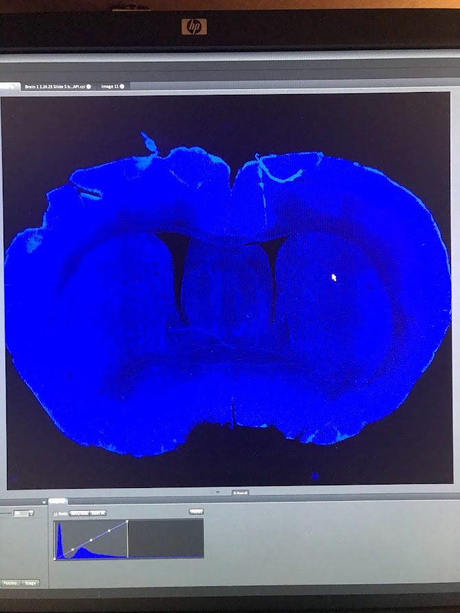
It was hard to describe the first time I saw this image. Yeah, there were some nicks and tears in the tissue (my hands were shaky during the perfusion), but this was something that I had wholly produced myself. And now, in front of me, was a snapshot of all that this mouse was.
See that little butterfly shaped area in the middle of the brain, right above the hole in the middle? That’s the hippocampus, where every memory is scrawled in your brain through a network of neurons. What mousy memories were kept in there? Looking at this picture, I can almost imagine what it would have been like in there when the mouse was alive. The holes in between and to the side of the “wings” are ventricles, gaps in the tissue where cerebrospinal fluid carrying nutrients and hormones was kept. I can almost imagine waves of it washing over the hippocampus, keeping this great curled machine firing, shooting sparks of electricity in patterns that say I am alive. I have lived. Here is what I have seen, heard, smelled, touched, tasted, hated, loved.
To be honest, this work can be kind of sad. After all, all life is precious. Sometimes, it can feel almost gruesome. To perfuse a mouse, you stick a needle of fixative into its beating heart, so it evenly diffuses and preserves the entire body. The night after I read this protocol, which had a video of this process in high definition, I felt almost too nervous to sleep. When I closed my eyes, I just saw that mouse heart beating out of its chest, beating, beating, beating…
The next morning, I admitted to my principal investigator that I wasn’t sure about trying out the perfusion that day. “Everything else, I’m okay with, but the way you can see the heart pumping freaks me out. I still want to try it, but…”
He nodded. “You never really feel comfortable with this part of the work.”
After a moment, he added, more seriously, “and that’s a good thing.”
Testing on animals is a controversial subject, and I can’t claim to have the definitive opinion. But, I think my experiences give me a special point of view. In the animal labs at this institute, the animals are well cared for. Until the last day they’re with us, we treat them with respect, care, and even love (what else do you call grown adults cooing at a 2 inch long mouse washing its tiny little face with its tiny little paws?). There’s laws, regulations, committees to ensure that animals are treated ethically. They’re given pain medications after surgeries, and during procedures (surgery and perfusions) they’re put under anesthesia. Once, our lab manager stayed in the surgery room with the mouse she was operating on until night fell to make sure she was there to care for him when he woke up.
And yet, we have to sacrifice these animals in the end, and we have complicated feelings about it. I think that’s good and necessary. If we feel sad at the animal’s death, we treat it well when it’s alive. If we feel a bit of horror at watching its life painlessly bleed away in the dissection pan, it means we placed a high value on that life in the first place. We try to perfect our surgical skills, write better protocols, and design better experiments to minimize mistakes and the number of mice who have to undergo this process. I would much prefer that than to become cold and callous.
Because the truth is, we don’t have a better process to study the brain in vivo. Treatments for neurodegenerative disorders, like Parkinson’s and Huntington’s (two diseases targeting the brain area our lab studies), mental illnesses, and mental disorders lag far behind those for many other parts of the body. And the simple reason is that we often can’t get into the nervous system to study it without causing permanent damage. Until better options arise, this is the best way to perform this life-saving research.
Whew. Things got really serious there. But working with something as big as life and death all the time makes you think about big questions. For me, ultimately this type of research is justifiable: this is the only way to validate that the research being conducted is working properly. It’s the only way to make sure that all the weird situations we put mice in weren’t done in vain. And getting to do every step of this process, going from a live mouse to a bright image on a screen–that’s something really special. Through that intricate picture, you can almost see something persisting, living on…helping us unlock the secrets of the brain. There is something to be said, here, about loving something by studying it: giving it all your attention while taking notes, appreciating the simple symmetry of the brain slice, using the gentlest touch to mount delicate tissue onto slides, admiring the natural structural variations that nature gifts every body and every brain.
Finally, I want you to think about your daily life, and the laboratory animals that made it possible. How many medications did you take today? What about cosmetics? Vaccines? Animal testing was where many of them began. One funny thing I’ve noticed recently is that researchers who work with “animals” tend to be the most adamant about using the term “humans and non-human animals”. Other species of animals are intertwined with us in inextricable ways. And there’s really no way to really convey my gratitude to a species that can not understand it.
I guess this article is my attempt to try.