I Got My Brain Scanned for Science

Yeah, I got my brain scanned for science. And you should too! (If you’re eligible, that is.)

Basically, I got an MRI scan of my brain done recently as part of a study at the GT/GSU Center for Advanced Brain Imaging (CABI). And while I was kind of nervous about the process, I wanted to write about the experience to help other people. If you’re about to get an MRI scan for medical reasons or for a study, I hope this article can relieve any fears or answer any questions you might have!

So, what is an MRI? MRI stands for Magnetic Resonance Imaging. An MRI machine looks like a very large tube with a donut sized hole on the end. During an MRI, you lay down in front of the donut and the technician moves you into the machine. By using magnets, the machine is able to take 3D images of your organs (in my case, my brain!). Since an MRI is so detailed and there’s no radiation risk, it is often used by doctors to detect tumors and look at brain injuries. There’s a version of an MRI called an fMRI, which shows which areas of your brain are activated. The f in fMRI stands for function, and fMRI images are able to help scientists understand how we think.

Since an MRI uses a really powerful magnet, you can’t bring anything metal in there. So, people with pacemakers, metal stents, braces, etc. might not be able to get an MRI. And when I say a powerful magnet, I mean it. The MRI machine at CABI is behind a heavy, access locked door with two large signs looking like the one on the right:
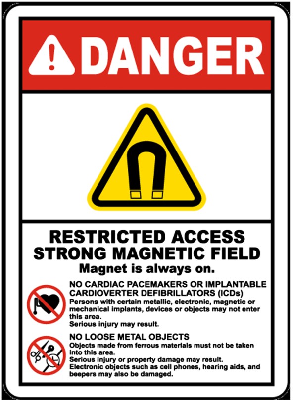
A danger sign with a red header saying danger. Underneath is a yellow triangle with a magnet symbol indicating a strong magnetic field.And if you have a morbid curiosity as to what would happen if you did bring in metal on or in you, check out this video: MRI vs Metal Coins (American Currency) DO NOT ATTEMPT To Replicate. Notice that just by being near the MRI, the camera goes all wonky. Some other metal things that you might not consider:

  • Some tattoo inks that include metal
  • Earrings
  • Hearing implants
  • Magnetic eyelashes
  • Those metal fasteners on pants

If you have any of these, it doesn’t mean you’re automatically disqualified from getting an MRI, but it’s important to bring it up to the technician or radiologist.

After I made sure I was metal free, I stepped into the MRI control room. There was a giant window where the researchers and technician would be able to see me laying in the MRI in the next room. I took off my jacket and shoes, feeling a bit jittery now. I was going to be INSIDE that giant thing.

The research assistant made her way over to me with a Vitamin E capsule and some tape. At my confused look, she explained, “Vitamin E shows up really brightly on the MRI, so we are gonna put this on your left forehead. That way, when the scans come in, we can tell which side is the left.” Makes sense to me. But that didn’t make it feel less weird when I put it on my head.

The technician led me into the MRI room, where I laid down on the extended platform. It felt like the kind of examination table they have at the doctor’s office, but it was more padded. That was to discourage people from moving around and messing up the scan. After I laid down, I felt the technician place down more padding around my head and feet. She also gave me earplugs and a bulb that I was supposed to squeeze if I needed anything once I was in the machine. As she did all this and wheeled me into the darkness of the machine, she told me what she was doing. This helped calm my nerves a lot, considering I couldn’t see what was going on. 

Finally, the technician turned the machine on, and I immediately appreciated the earplugs. The beeping reminded me of an alarm clock. I heard that some people can fall asleep during an MRI, but that wasn’t me. It wasn’t earsplitting, but just loud enough that it never faded into the background.

My earplugs were attached to an earpiece. After a burst of static, I heard the research assistant over the beeps. 

“Okay, we’re going to do the first part of the MRI and take a picture of your brain’s structure. Do you feel okay?”

I nodded. There was a pause. “Wait, sorry,” I said, smiling sheepishly to myself in the dark of the machine. Obviously, they couldn’t see me. “Yes, I’m all ready.”

The actual imaging itself was boring, but I passed the time imagining the magnetic beam hitting all of my cells. After a while, I could almost feel the beam on my skin. There wasn’t actually anything, of course. Then, the fMRI was done. The beeping was a different rhythm–more pleasant, in my opinion.

While the scan was being performed, I completed a memory task as part of the study. A screen was put in front of me while I was still in the scanner, with the image projected from a computer screen in the control room using some mirrors. Using a control pad of buttons, I was even able to manipulate what was on the screen. I didn’t even know you could do all that while in an MRI machine!

Soon, I was able to be released from the dark void I had been in previously. The whole process took around half an hour–much shorter than I expected. Other than some numbness from staying as still as possible, I felt alright. And I was excited to see what my brain really looked like!
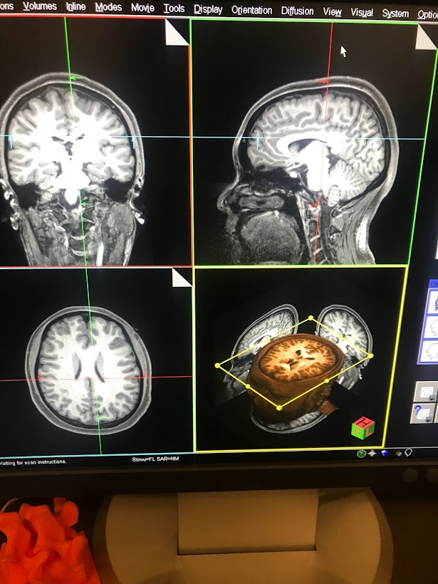
A picture of my MRI scan from the coronal, sagittal, and horizontal views.
This is my brain! From the top left going counterclockwise, that is the coronal view, horizontal view, and sagittal view. The bottom right shows all the slices of my brain that were taken, which you can see by moving the yellow lines.

And there it was–all of my consciousness, my memories, my thoughts, my emotions, dreams, fears all up on that screen. It looked so complicated and fragile and beautiful. As a neuroscience major, I saw pictures of brains every day. But nothing hits quite like seeing your own brain up there in black and white. And there I was, using my brain to understand that what I saw in front of me was itself. It’s such an indescribable feeling!

And that reason, beyond just the contributions to science, is why I recommend getting an MRI done as part of a study. After this experience, I felt myself thinking about my own brain with more affection. This squishy, bumpy organ is me, and I should treat it with the study and love that it deserves.


References

https://www.researchgate.net/figure/Magnetic-field-warning-sign_fig1_345758192

Your journey starts here! Let’s talk!

ThemeRex © 2026. All rights reserved.
  Timothy Durham was convicted of rape when a test showed his DNA genotype matched the DNA recovered from the crime scene. Eye witnesses testified that Durham was in a different state at the time of the incident, but he wasn’t exonerated from his 3000 year sentence until follow up DNA analysis showed that the preliminary forensic analysis used in the trial was misinterpreted (Thomas et al.). DNA testing serves an invaluable part in our criminal justice system, but it comes with complications and bias that can lead to wrongful convictions and misinformed juries.  

Background:

Forensic genetics aims to identify the origin of a biological sample. It can be traced back to Karl Landsteiner who discovered human ABO blood groups. With blood groups, suspects could be eliminated based on their blood type. For example, if a suspect had type A blood and there was type B blood left at the scene by the perpetrator, the investigators could rule out the suspect. Unfortunately, the reverse didn’t work. If the suspect and perpetrator both had the same blood type, it can’t be concluded that the suspect was guilty. Blood identifiers evolved into 8 distinct categories which improved the ability to eliminate suspects, but could only conclude that (if the system matched) it might be the perpetrator or this person might be related to the perpetrator (Jobling and Gill). The field evolved in the 1980s with the discovery of Minisatellites, lines of repetitive DNA that don’t encode proteins. Minisatellite patterns in DNA create what we know today as the DNA fingerprint, and it has a much higher accuracy rate than previous methods (Augustyn et al.). Around the same time, a breakthrough for victims of sexual assault was discovered. In the case of assault, bodily fluids of both the victim and the assaulter can get mixed. A process called differential lysis was created which could selectively increase the concentration of sperm in a fluid sample thereby allowing for accurate DNA testing (Jobling and Gill). Forensics evolved to use single-locus probe testing after several years because of its ease of interpretation. It looked at specific minisatellites that had high variability between subjects. This was the first DNA-based method to be used in a criminal investigation. Two murder homicides from several years apart were connected using DNA evidence. The evidence was then used to exonerate an innocent man, and the guilty party was found when he tried to evade mandatory DNA testing for all men in the small town. His guilt was then reinforced when the DNA profiles matched, and he gave a confession (Jobling and Gill). Finding DNA at a crime scene doesn’t guarantee that you’ll find a murderer. There are issues with non-pure samples and limited samples that make DNA work difficult, and accreditation of facilities and new techniques and take time (Jobling and Gill).  

Subjectivity of DNA Interpretation:

In a 2009 publication, the National Academy of Science discusses the possibility for subjective influence in lab based forensics. The interpretation bias happens in complex cases where, for example, there could be a mixture of DNA. A study published in 2011 looked specifically at this issue. A gang rape happened in Georgia, and the mixture in DNA from the case was used to prosecute one suspect after another suspect took a plea bargain to testify against him. With DNA mixtures, there are three possible conclusions: suspect excluded, suspect cannot be excluded, and inconclusive. If the suspect is excluded, it means that there’s little to no possibility that his DNA is part of the sample. If the suspect cannot be excluded, it means that there’s a possibility that his DNA is part of the sample. In the trial, the DNA examiners who knew the context of the case concluded that the suspect being prosecuted could not be excluded. The 2011 study gave the DNA evidence from this case and the DNA profiles of the suspects to 17 forensic examiners. These examiners had no context of the case, and they all worked independently. Of the 17, only 1 came to the same conclusion (suspect cannot be excluded) as the examiners who knew the context of the case. Twelve of the 17 concluded that the suspect who was prosecuted and convicted had no relation to the DNA found in the sample. Not only does this show that subjectivity occurs due to the context of the case, but the 17 examiners came up with different findings based on their past experiences meaning that not all DNA analysis is objective. Some organizations like the Scientific Working Group on DNA Analysis Methods say that quantitative statistical evidence should be presented with every categorical conclusion, but the US doesn’t require this (Dror and Hampikian).

False Positives:

There are two categories that cause wrongful identification in a DNA report: coincidental matches and false positives. Coincidental matches are DNA matches between two unrelated people. The probability of these matches is about 1 to 500,000 for white individuals and about 1 to 350,000 for black individuals. This small probability of coincidental matches makes people feel confident in DNA evidence. False positives, on the other hand, are matches between DNA and test DNA that result from issues in the forensics lab. There have been multiple cases of forensics labs accidentally switching victim and suspected rapist DNA which lead to convictions that were later overturned when the lab realized the error. There are also cases of misreading or misunderstanding a sample. In a case from 1993, a man was convicted of rape because someone interpreted a mixed sample of victim and perpetrator DNA as an individual sample. The conviction was again overturned. To prevent false convictions, retesting DNA is allowed, but it’s not an efficient solution. Many people are pushing for statistical information on the probability of false positives, so that the jurors can make accurate judgements based on DNA evidence (Thomas et al.).  

Conclusion:

DNA analysis is a useful tool for the criminal justice system, but subjectivity can be an issue in forensic testing. To combat wrongful convictions, there are organizations like The Innocence Project who use DNA testing to free the innocent, but reformed forensic testing standards should be put in place to prevent subjectivity and to ensure juries are correctly informed during trials. https://www.innocenceproject.org/    

Citation:

Augustyn , Adam, et al. “DNA Finerprinting.” Encyclopedia Britannica , 26 Apr. 2018, www.britannica.com/science/DNA-fingerprinting#ref153186. Dror, Itiel E., and Greg Hampikian. “Subjectivity and Bias in Forensic DNA Mixture Interpretation.” Science & Justice, vol. 51, no. 4, 2011, pp. 204–208., doi:10.1016/j.scijus.2011.08.004. Jobling, Mark A., and Peter Gill. “Encoded Evidence: DNA in Forensic Analysis.” Nature Reviews: Genetics, vol. 5, no. 10, Oct. 2004, pp. 739–751., doi:10.1038/nrg1455. Thompson, William C, et al. “How the Probability of a False Positive Affects the Value of DNA Evidence.” Journal of Forensic Sciences, vol. 48, no. 1, Jan. 2003, pp. 1–8., doi:10.1520/jfs2001171.