How to make glow in the dark cells

While these cells don’t technically glow in the dark, scientists have found ways to make cells fluorescent which means they glow when exposed to radiation such as UV light. When a cell has specific ions or proteins fluorescent proteins bind to these small molecules and make the cell appear as if it’s glowing. 

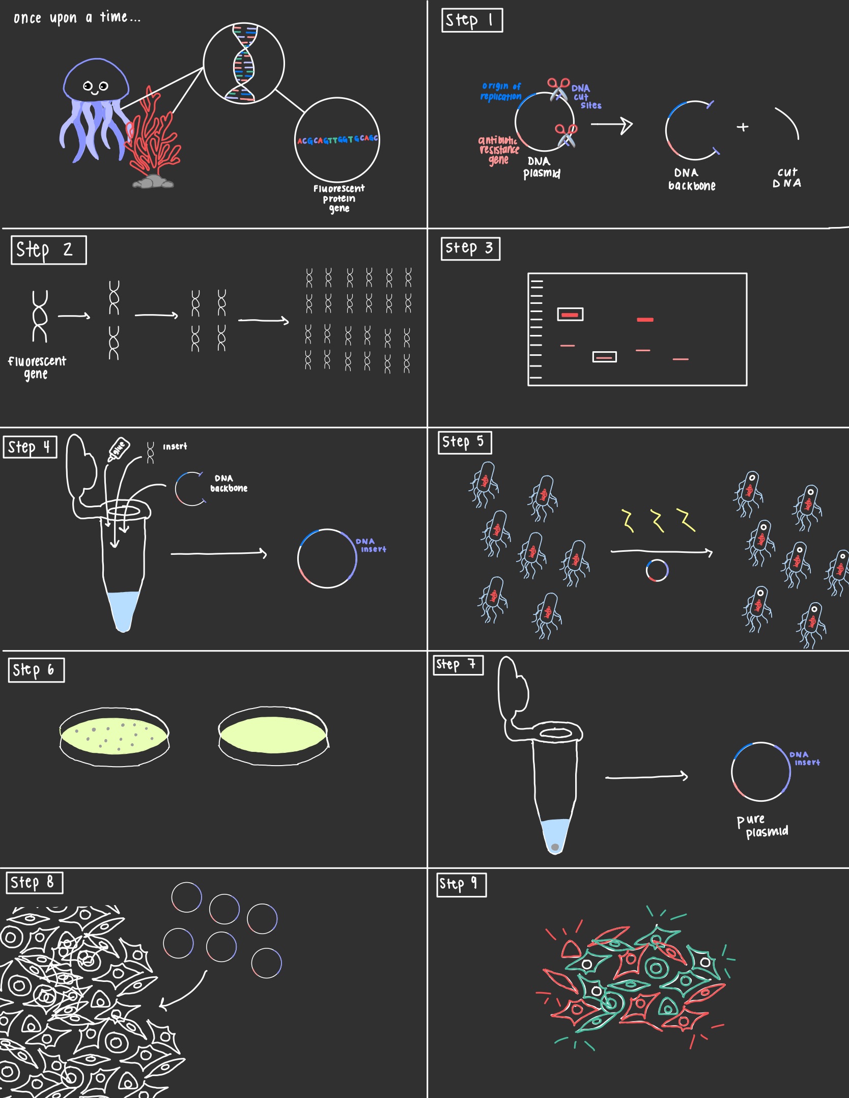
These special proteins were first discovered in animals like jellyfish and plants like coral. Scientists were able to isolate the genes that were responsible for producing the fluorescent proteins and using a technique called molecular cloning can insert the DNA into our cells of choice. 

Step 1: The first step is to choose a plasmid, a circular piece of DNA found in bacteria. The plasmid chosen must have an origin of replication where DNA replication begins, a gene for antibiotic resistance, and DNA cut sites. Restriction enzymes are proteins that act as molecular scissors and cut the plasmid at specific cut sites. This turns the circular plasmid DNA into a linear piece of DNA. 

Step 2: This isn’t always necessary, but the fluorescent gene can be amplified using a technique known as polymerase chain reaction. In a PCR the DNA undergoes cycles of denaturation, annealing, and elongation where the DNA is first turned into single stranded DNA, then the primer tells the DNA where to start extending, and then finally the DNA is extended. At the end of each cycle the amount of DNA is doubled leading to millions of copies of DNA within an hour. 

Step 3: By this point, we have our amplified PCR DNA and our plasmid DNA that has been cut to produce the backbone and pieces of cut DNA. In order to separate the backbone plasmid from the cut piece we will use gel electrophoresis. A gel electrophoresis separates DNA by size by passing an electric current through a gel containing DNA. Because DNA is negatively charged it will migrate towards the positively charged terminal of the gel with the smaller pieces traveling further than the larger pieces. Because our backbone is larger than the cut DNA, when the pieces separate we can cut out the band closer to the top, and purify it to get rid of the gel leaving just the DNA. 

Step 4: It’s finally time to glue our DNA pieces together, and the key player is a protein known as DNA ligase. DNA ligase will be added to a tube with our DNA backbone and insert. Following a ligation reaction a plasmid will be re-formed that contains our original backbone with the fluorescent gene inserted in. 

Step 5 and Step 6: The next two steps are crucial for ensuring that the final product is pure plasmid. This is important because the DNA ligase can make mistakes and may glue backbones together, fail to add in the insert, etc. In step 5 the plasmid is inserted into bacterial cells, usually e-coli cells through a heat shock. Once the bacteria take up the plasmid they are plated onto agar plates containing antibiotics and allowed to grow. Remember the three things every plasmid needs to include (hint: it’s an antibiotic resistance gene)? This is because when the cells are plated only those that have a fully formed plasmid will be able to grow. 

Step 7: Now we can select a bacterial colony from our plate and using a process known as miniprep isolate the plasmid DNA from these cells. 

Step 8: The plasmid DNA can then be introduced into your cells of choice. There are many techniques to accomplish this but a common one is called calcium phosphate transfection. 

Step 9: TA-DA! Your cells should now glow under the right conditions! 

Sources:

Ashwini, M., Murugan, S.B., Balamurugan, S. et al. Advances in molecular cloning. Mol Biol 50, 1–6 (2016). https://doi.org/10.1134/S0026893316010131

Tsien RY. The green fluorescent protein. Annu Rev Biochem. 1998;67:509-44. doi: 10.1146/annurev.biochem.67.1.509. PMID: 9759496.
cover image created using canva

Your journey starts here! Let’s talk!

ThemeRex © 2026. All rights reserved.
  Timothy Durham was convicted of rape when a test showed his DNA genotype matched the DNA recovered from the crime scene. Eye witnesses testified that Durham was in a different state at the time of the incident, but he wasn’t exonerated from his 3000 year sentence until follow up DNA analysis showed that the preliminary forensic analysis used in the trial was misinterpreted (Thomas et al.). DNA testing serves an invaluable part in our criminal justice system, but it comes with complications and bias that can lead to wrongful convictions and misinformed juries.  

Background:

Forensic genetics aims to identify the origin of a biological sample. It can be traced back to Karl Landsteiner who discovered human ABO blood groups. With blood groups, suspects could be eliminated based on their blood type. For example, if a suspect had type A blood and there was type B blood left at the scene by the perpetrator, the investigators could rule out the suspect. Unfortunately, the reverse didn’t work. If the suspect and perpetrator both had the same blood type, it can’t be concluded that the suspect was guilty. Blood identifiers evolved into 8 distinct categories which improved the ability to eliminate suspects, but could only conclude that (if the system matched) it might be the perpetrator or this person might be related to the perpetrator (Jobling and Gill). The field evolved in the 1980s with the discovery of Minisatellites, lines of repetitive DNA that don’t encode proteins. Minisatellite patterns in DNA create what we know today as the DNA fingerprint, and it has a much higher accuracy rate than previous methods (Augustyn et al.). Around the same time, a breakthrough for victims of sexual assault was discovered. In the case of assault, bodily fluids of both the victim and the assaulter can get mixed. A process called differential lysis was created which could selectively increase the concentration of sperm in a fluid sample thereby allowing for accurate DNA testing (Jobling and Gill). Forensics evolved to use single-locus probe testing after several years because of its ease of interpretation. It looked at specific minisatellites that had high variability between subjects. This was the first DNA-based method to be used in a criminal investigation. Two murder homicides from several years apart were connected using DNA evidence. The evidence was then used to exonerate an innocent man, and the guilty party was found when he tried to evade mandatory DNA testing for all men in the small town. His guilt was then reinforced when the DNA profiles matched, and he gave a confession (Jobling and Gill). Finding DNA at a crime scene doesn’t guarantee that you’ll find a murderer. There are issues with non-pure samples and limited samples that make DNA work difficult, and accreditation of facilities and new techniques and take time (Jobling and Gill).  

Subjectivity of DNA Interpretation:

In a 2009 publication, the National Academy of Science discusses the possibility for subjective influence in lab based forensics. The interpretation bias happens in complex cases where, for example, there could be a mixture of DNA. A study published in 2011 looked specifically at this issue. A gang rape happened in Georgia, and the mixture in DNA from the case was used to prosecute one suspect after another suspect took a plea bargain to testify against him. With DNA mixtures, there are three possible conclusions: suspect excluded, suspect cannot be excluded, and inconclusive. If the suspect is excluded, it means that there’s little to no possibility that his DNA is part of the sample. If the suspect cannot be excluded, it means that there’s a possibility that his DNA is part of the sample. In the trial, the DNA examiners who knew the context of the case concluded that the suspect being prosecuted could not be excluded. The 2011 study gave the DNA evidence from this case and the DNA profiles of the suspects to 17 forensic examiners. These examiners had no context of the case, and they all worked independently. Of the 17, only 1 came to the same conclusion (suspect cannot be excluded) as the examiners who knew the context of the case. Twelve of the 17 concluded that the suspect who was prosecuted and convicted had no relation to the DNA found in the sample. Not only does this show that subjectivity occurs due to the context of the case, but the 17 examiners came up with different findings based on their past experiences meaning that not all DNA analysis is objective. Some organizations like the Scientific Working Group on DNA Analysis Methods say that quantitative statistical evidence should be presented with every categorical conclusion, but the US doesn’t require this (Dror and Hampikian).

False Positives:

There are two categories that cause wrongful identification in a DNA report: coincidental matches and false positives. Coincidental matches are DNA matches between two unrelated people. The probability of these matches is about 1 to 500,000 for white individuals and about 1 to 350,000 for black individuals. This small probability of coincidental matches makes people feel confident in DNA evidence. False positives, on the other hand, are matches between DNA and test DNA that result from issues in the forensics lab. There have been multiple cases of forensics labs accidentally switching victim and suspected rapist DNA which lead to convictions that were later overturned when the lab realized the error. There are also cases of misreading or misunderstanding a sample. In a case from 1993, a man was convicted of rape because someone interpreted a mixed sample of victim and perpetrator DNA as an individual sample. The conviction was again overturned. To prevent false convictions, retesting DNA is allowed, but it’s not an efficient solution. Many people are pushing for statistical information on the probability of false positives, so that the jurors can make accurate judgements based on DNA evidence (Thomas et al.).  

Conclusion:

DNA analysis is a useful tool for the criminal justice system, but subjectivity can be an issue in forensic testing. To combat wrongful convictions, there are organizations like The Innocence Project who use DNA testing to free the innocent, but reformed forensic testing standards should be put in place to prevent subjectivity and to ensure juries are correctly informed during trials. https://www.innocenceproject.org/    

Citation:

Augustyn , Adam, et al. “DNA Finerprinting.” Encyclopedia Britannica , 26 Apr. 2018, www.britannica.com/science/DNA-fingerprinting#ref153186. Dror, Itiel E., and Greg Hampikian. “Subjectivity and Bias in Forensic DNA Mixture Interpretation.” Science & Justice, vol. 51, no. 4, 2011, pp. 204–208., doi:10.1016/j.scijus.2011.08.004. Jobling, Mark A., and Peter Gill. “Encoded Evidence: DNA in Forensic Analysis.” Nature Reviews: Genetics, vol. 5, no. 10, Oct. 2004, pp. 739–751., doi:10.1038/nrg1455. Thompson, William C, et al. “How the Probability of a False Positive Affects the Value of DNA Evidence.” Journal of Forensic Sciences, vol. 48, no. 1, Jan. 2003, pp. 1–8., doi:10.1520/jfs2001171.